Germ Cell Sorter
Designed and built end to end, from the chip to the control loop
For my MEng final-year project I designed and built a complete microfluidic fluorescence-activated cell sorter (µFACS), on my own, from the chip up. The primordial germ cells that fertility research depends on are rare and fragile, and the commercial machines that sort them cost $80,000 to $150,000, run on complex protocols, and shear delicate cells to death. My sorter set out to do it gently and cheaply: focus the cells in a custom chip, single out the GFP-tagged germ cells with a bespoke dual-channel optical system, and sort them with a soft pressure pulse fired by real-time machine vision. I built every subsystem myself, the microfluidics, the Zemax-optimised optics, the electronics, and the LabVIEW control, and got each one working.
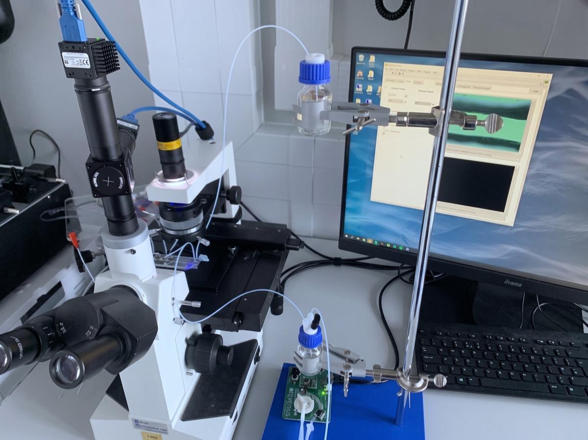
The rig

What it does
- Built end to end, solo, as a final-year undergraduate: the microfluidic chip, the optics, the electronics, and the real-time control software.
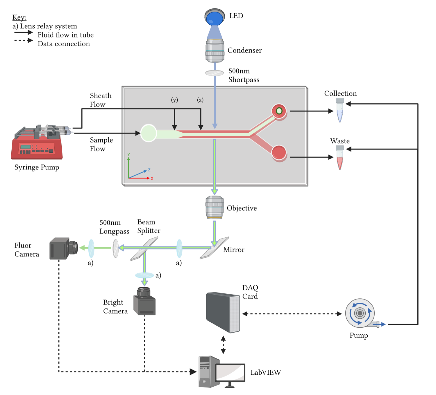
- Focuses fragile cells gently in a custom laser-cut chip using 3D hydrodynamic focusing, shrinking the imaging region while keeping shear low.
- Images brightfield and fluorescence at once through a 4f telecentric relay I optimised in Zemax and mounted via a 3D-printed, lathe-tapped adapter, so GFP-tagged germ cells stand out from everything else.
- Closes the loop in LabVIEW: real-time machine vision confirms a target, then a DAQ-driven pressure pulse redirects it into the collection outlet.
The problem
Primordial germ cells are central to studying the germline and to fertility research, but they are rare, fragile, and arrive mixed with somatic cells that overgrow them in culture. The two cell types are too similar in size for passive methods like filters to tell apart, so sorting them needs active detection of each cell. The catch is that the machines built for that, industry-standard flow cytometers, are costly, run on complex protocols, and are rough enough on cells to kill the fragile ones. The goal was to sort the germ cells actively but gently, at a fraction of the cost and complexity.
What it demonstrated
Tested with 10 µm fluorescent and transparent microspheres as cell surrogates, each subsystem did its job: the chip focused the sample stream, the optics cleanly told fluorescent targets from transparent particles, and a pressure pulse redirected the central streamline into the collection branch on cue. It stopped short of a production sorter, quantifying sort purity and yield was the clear next step, and another student later carried the chip design and sorting evaluation forward. As a solo final-year project spanning microfluidics, custom optics, electronics, machine vision, and real-time control, it is where I learned to take an instrument from a blank chip to a working loop.
A closer look


